Expert Keratoconus Care

Keratoconus is a progressive corneal condition that causes blurred, distorted vision, glare, halos, and increasing light sensitivity. It occurs when the cornea thins and bulges into a cone-like shape, making it harder for light to focus clearly on the retina.1

At Innovative Eye Care, keratoconus diagnosis and management is a core clinical focus. Our optometrists use advanced corneal imaging to detect early changes, monitor progression, improve vision with specialty contact lenses, and refer for surgical treatment when needed.

Our principal optometrist, Lachlan Hoy, authored the keratoconus chapter in the textbook Contact Lenses (2019), reflecting our focus on evidence-based keratoconus care.
Book a keratoconus assessment
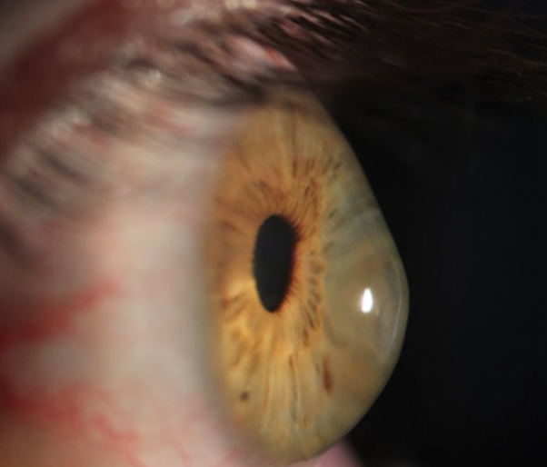
Side view slit lamp image showing advanced keratoconus with cone-shaped cornea

What Is Keratoconus?

Keratoconus affects the cornea, the clear front surface of the eye. As the cornea becomes thinner and more irregular, vision can become distorted and difficult to correct with standard glasses.1,2

Common symptoms include:

  • Blurred or distorted vision
  • Halos and glare, especially at night
  • Increased light sensitivity
  • Frequent prescription changes
  • Increasing astigmatism
  • Poor vision even with glasses

Symptoms often begin in the teenage years and may continue progressing into the 30s or 40s.2,3 In Australia, studies suggest keratoconus affects around 1 in 84 young adults, making early detection important.4

Man rubbing his eye, a risk factor for keratoconus progression

Keratoconus Risk Factors

Keratoconus can involve both genetic and environmental factors.2,3

Risk factors include:

  • Family history of keratoconus
  • Allergies, hay fever, asthma, or eczema
  • Chronic eye rubbing
  • Down syndrome
  • Marfan syndrome
  • Ehlers-Danlos syndrome
  • Floppy eyelid syndrome

If you have these risk factors and notice frequent prescription changes, night glare, or worsening vision, book a comprehensive assessment.
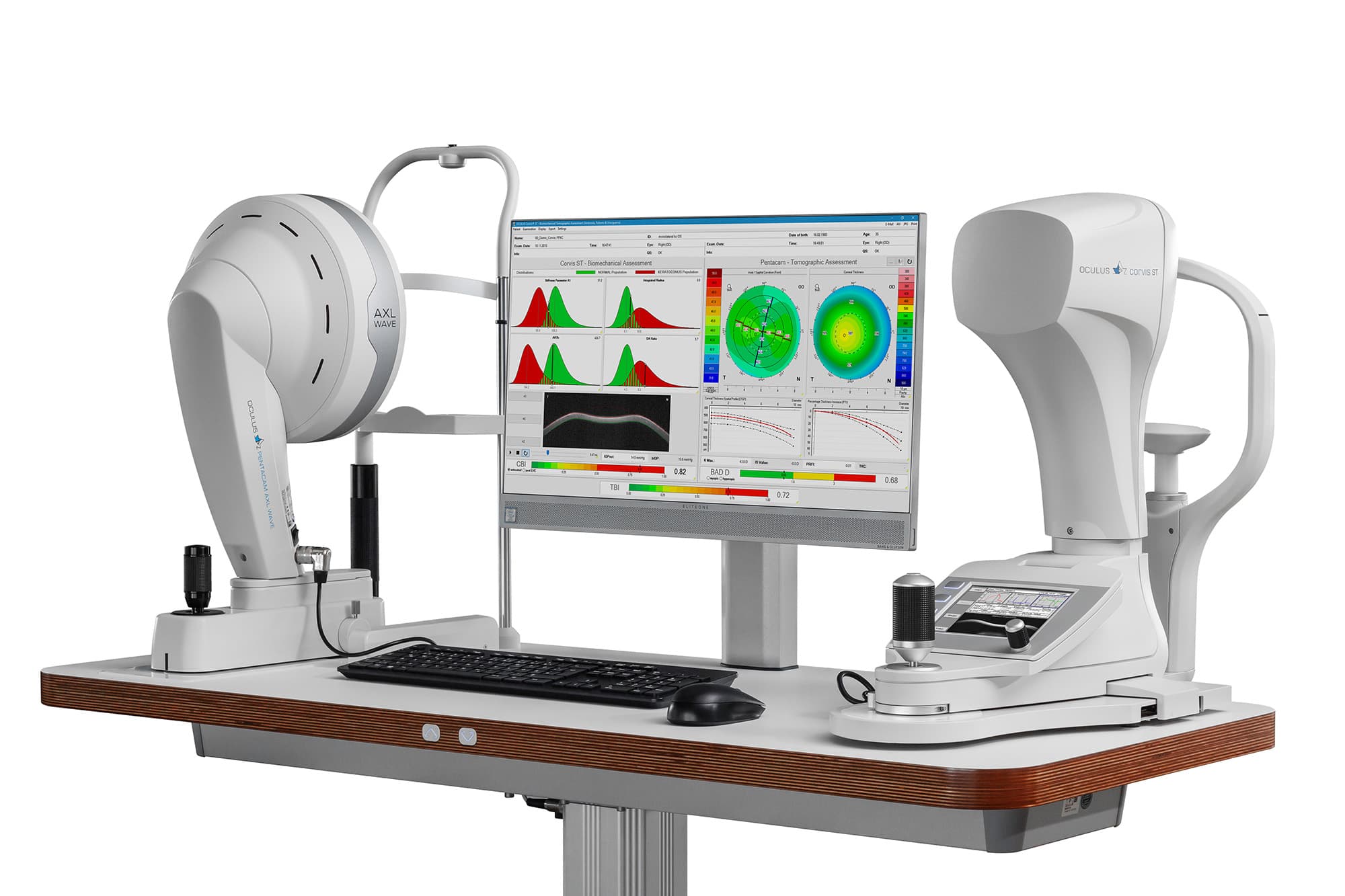
Pentacam AXL Wave and Corvis ST used for keratoconus diagnosis at Innovative Eye Care Adelaide

Early Keratoconus Diagnosis

Early diagnosis helps protect long-term vision. Keratoconus can progress before symptoms feel severe, so advanced imaging is essential.5,6

At Innovative Eye Care, we use:

  • Corneal Tomography – Gold-standard 3D mapping of corneal shape and thickness
  • Corvis ST – Measures corneal biomechanics for early detection
  • Wavefront Aberrometry – Assesses higher-order optical distortions
  • Corneal Topography – Detailed surface mapping
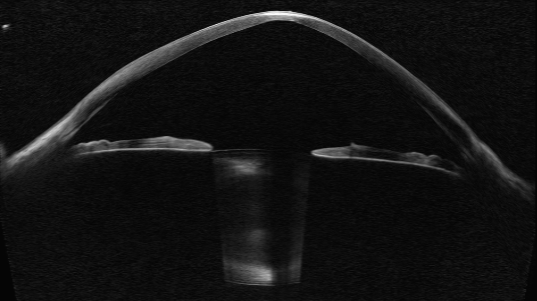
  • Anterior OCT – Cross-sectional corneal imaging
  • Slit Lamp Examination – Clinical assessment of corneal structure

These technologies allow us to detect subtle changes, long before they become visually significant.
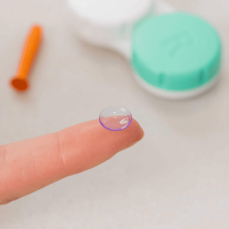
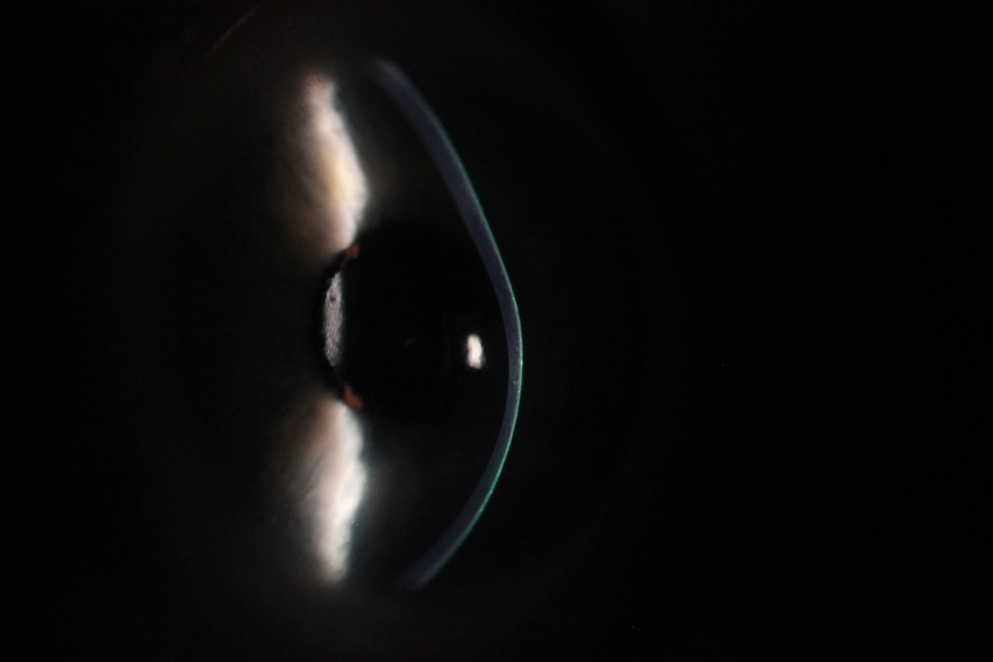
Rigid contact lens for keratoconus vision correction

Keratoconus Treatment and Management Options

Keratoconus management depends on severity, progression, age, vision needs, and corneal health.

Avoid Eye Rubbing

Eye rubbing is linked with keratoconus progression. Managing allergies and reducing rubbing can help reduce risk.7

Glasses and Soft Contact Lenses

In early keratoconus, glasses or soft contact lenses may provide clear vision. As the cornea becomes more irregular, these options often become less effective.1,3

Rigid Gas Permeable Contact Lenses

Rigid gas permeable lenses can improve vision by creating a smoother optical surface over the irregular cornea.3
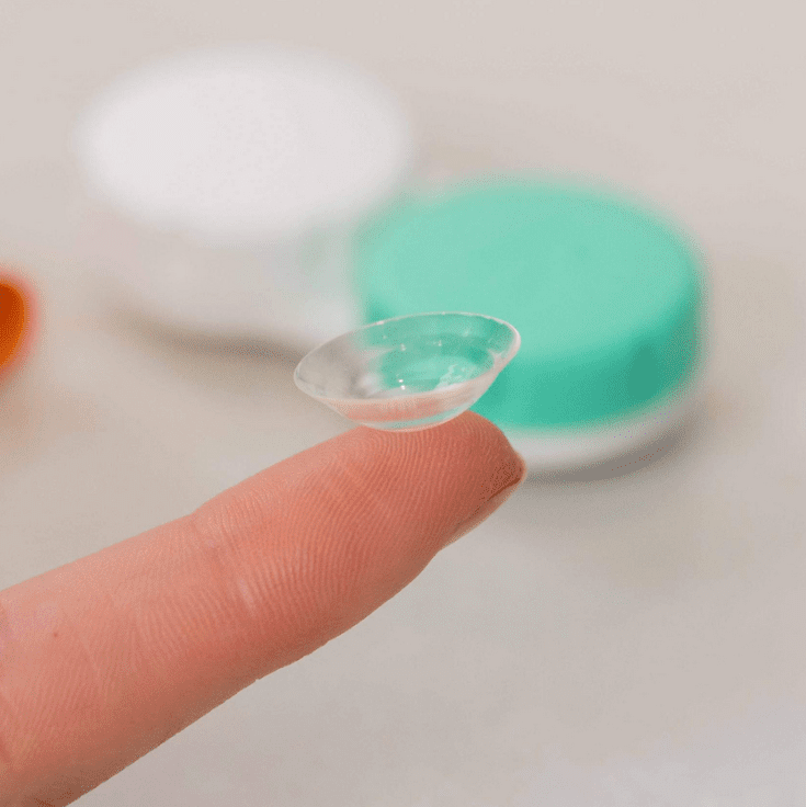
Scleral contact lens for keratoconus vision correction

Scleral Lenses for Keratoconus

Scleral lenses are often used for moderate to advanced keratoconus. They vault over the cornea and rest on the white part of the eye, creating a smooth optical surface that can improve clarity and comfort.3

Our team uses EyeSpace fitting software to design precise, comfortable lenses. Poorly fitted lenses may worsen corneal stress, so expert fitting and ongoing review are important.8,9

Keratocous Contact Lenses
Royal Adelaide Hospital for specialist ophthalmology care and keratoconus referral in Adelaide

Surgical Options for Keratoconus (In Collaboration with Ophthalmology)

Corneal Collagen Cross-Linking

Corneal collagen cross-linking, or CXL, aims to strengthen the cornea and slow or stop progression.10 It works best when progressive keratoconus is detected early. The introduction of CXL has been associated with a reduction in corneal transplantation for keratoconus, supporting its role in reducing progression to advanced disease.11

Intracorneal Ring Segments

Intracorneal ring segments may reshape the cornea in selected mild to moderate cases.3

Corneal Transplant

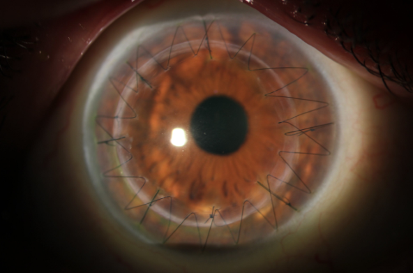
Corneal transplant, or keratoplasty, is usually reserved for advanced keratoconus with severe scarring, poor lens tolerance, or vision that cannot be corrected with lenses.1,3
Keratoconus vs Pellucid Marginal Degeneration

Corneal graft after keratoconus surgery in Adelaide

Pellucid marginal degeneration, or PMD, is a rarer corneal thinning condition that can look similar to keratoconus. It usually affects the lower cornea and can cause high against-the-rule astigmatism.5 Accurate diagnosis matters because lens fitting and management can differ.

Innovative Eye Care optometrist explaining corneal scan results during keratoconus consultation in Adelaide

When to Book a Keratoconus Assessment

Book an assessment if you notice:

  • Increasing astigmatism
  • Frequent prescription changes
  • Blurred or distorted vision
  • Glare or halos at night
  • Light sensitivity
  • Vision that glasses no longer correct clearly
  • Family history of keratoconus
  • Early assessment can help protect your long-term vision.
Book a keratoconus assessment in Adelaide or Henley Beach

References

  1. Lawless M, Coster DJ, Phillips AJ, Loane M. Keratoconus: diagnosis and management. Aust N Z J Ophthalmol. 1989;17(1):33-60. https://pubmed.ncbi.nlm.nih.gov/2527524/
  2. Rabinowitz YS. Keratoconus. Surv Ophthalmol. 1998;42(4):297-319. doi:10.1016/s0039-6257(97)00119-7
  3. Romero-Jiménez M, Santodomingo-Rubido J, Wolffsohn JS. Keratoconus: a review. Cont Lens Anterior Eye. 2010;33(4):157-166; quiz 205. doi:10.1016/j.clae.2010.04.006
  4. Chan E, Chong EW, Lingham G, et al. Prevalence of Keratoconus Based on Scheimpflug Imaging: The Raine Study. Ophthalmology. 2021;128(4):515-521. doi:10.1016/j.ophtha.2020.08.020
  5. Gomes JAP, Rapuano CJ, Belin MW, Ambrósio R, Group of Panelists for the Global Delphi Panel of Keratoconus and Ectatic Diseases. Global Consensus on Keratoconus Diagnosis. Cornea. 2015;34(12):e38-39. doi:10.1097/ICO.0000000000000623
  6. Li X, Rabinowitz YS, Rasheed K, Yang H. Longitudinal study of the normal eyes in unilateral keratoconus patients. Ophthalmology. 2004;111(3):440-446. doi:10.1016/j.ophtha.2003.06.020
  7. Balasubramanian SA, Pye DC, Willcox MDP. Effects of eye rubbing on the levels of protease, protease activity and cytokines in tears: relevance in keratoconus. Clin Exp Optom. 2013;96(2):214-218. doi:10.1111/cxo.12038
  8. Macsai MS, Varley GA, Krachmer JH. Development of keratoconus after contact lens wear. Patient characteristics. Arch Ophthalmol. 1990;108(4):534-538. doi:10.1001/archopht.1990.01070060082054
  9. Korb DR, Finnemore VM, Herman JP. Apical changes and scarring in keratoconus as related to contact lens fitting techniques. J Am Optom Assoc. 1982;53(3):199-205.
  10. Galvis V, Tello A, Ortiz AI, Escaf LC. Patient selection for corneal collagen cross-linking: an updated review. Clin Ophthalmol. 2017;11:657-668. doi:10.2147/OPTH.S101386
  11. Godefrooij DA, Gans R, Imhof SM, Wisse RPL. Nationwide reduction in the number of corneal transplantations for keratoconus following the implementation of cross-linking. Acta Ophthalmol. 2016;94(7):675-678. doi:10.1111/aos.13095

Speak to our friendly team today

Book your appointment now for personalised eye care tailored just for you.