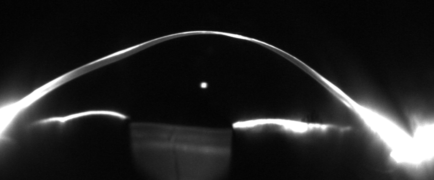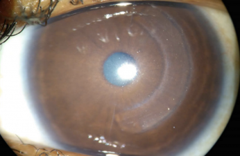
A patient with keratoconus recently returned to Innovative Eye Care approximately five weeks after undergoing corneal allogenic intrastromal ring segment implantation, or CAIRS, in Queensland. Historically, her best spectacle-corrected visual acuity was 6/12, with approximately 6/10 achieved in a scleral contact lens.
Our initial concern was whether surgical reshaping would improve her optical quality or introduce additional aberrations into an already irregular cornea. At this early review, the outcome was encouraging. Spectacle-corrected acuity had improved to 6/6, measured higher-order aberrations had approximately halved, and the patient reported substantially better vision. Her only reported visual disturbance was occasional dysphotopsia.
Pre-CAIRS
- Unaided Vision: 6/60+
- Refraction and best corrected spectacle lens acuity: +4.50/-7.75x86 (6/15)
Post-CAIRS
- Unaided Vision: 6/20-
- Refraction and best corrected spectacle lens acuity: +3.50/-5.75x43 (6/6-)
What is CAIRS?
CAIRS involves implanting segments of donor corneal stromal tissue into intrastromal channels. The aim is to reshape the ectatic cornea, flatten the cone and improve visual function. The original clinical report described CAIRS combined with corneal cross-linking in 24 eyes, with improvements in visual acuity and topographic parameters over 6 to 18 months (Jacob et al., 2018).
For optometrists, the relevant question is how this reshaping changes the patient's usable vision, spectacle correction and contact lens requirements.
Tomography Findings
The right-eye Pentacam measurement comparison shows a substantial redistribution of anterior corneal curvature. The previously steep central and paracentral region has flattened, with increased curvature in surrounding areas.
Displayed Kmax decreased from 61.9 D to 55.4 D, a reduction of approximately 6.5 D. Mean anterior keratometry decreased from 50.6 D to 46.5 D, and anterior corneal astigmatism decreased from 10.4 D to 7.0 D. The tangential difference map shows localised flattening exceeding 10 D within the previously steep region.
These findings illustrate why Kmax alone cannot fully describe the postoperative result. The location and distribution of curvature changes also matter when assessing the optical zone.
.png)
Aberrometry Findings
Both supplied aberration analyses use a 5.0 mm analysis diameter, allowing a more meaningful comparison than measurements taken over different apertures.
Without the scleral lens, higher-order aberration root mean square, or HOA RMS, decreased from 2.103 µm to 1.041 µm, approximately a 50% reduction. The displayed coma magnitude decreased from 1.602 µm to 0.583 µm, approximately a 64% reduction.
.png)
.png)
Visual Function and Dysphotopsia
The improvement from historical spectacle-corrected acuity of 6/12 to 6/6 was the most clinically meaningful finding. The patient's report of substantially better vision supported the measured outcome.
Her occasional dysphotopsia also deserves continued assessment. Achieving 6/6 does not capture every aspect of visual quality. Follow-up should document the symptom's frequency, severity and effect on activities such as night driving, alongside acuity and refraction.
How does this fit with the evidence?
A 2026 retrospective study of 25 eyes with advanced keratoconus reported improvements in visual acuity, refraction and keratometry after CAIRS, with a significant reduction in coma. Total HOA reduction did not reach statistical significance, and spherical aberration increased. These findings support assessing individual aberration terms alongside total RMS. The study's small sample, retrospective design and mean follow-up of approximately 11 months limit conclusions about predictability and durability (Yesilirmak et al., 2026).
This patient's five-week result is an encouraging early outcome.
Implications for Optometric Follow-Up
Post-CAIRS review should combine refraction, corrected acuity, repeatable tomography, matched-aperture aberration analysis and the patient's experience of vision.
For an existing scleral lens wearer, the altered corneal geometry warrants reassessment of clearance, fit and over-refraction before continued use. The historical 6/10 scleral acuity cannot establish the lens's postoperative performance.
Optical rehabilitation and progression management also require separate assessment. Early flattening does not demonstrate that ectasia has stabilised. Cross-linking has evidence for treating progressive keratoconus, and ongoing care should reflect the patient's documented treatment history and the operating surgeon's plan (Wittig-Silva et al., 2014).
This case demonstrates the value of looking beyond a flatter map. At five weeks, improved spectacle acuity, reduced HOA RMS and the patient's subjective improvement together provide a stronger account of the outcome than keratometry alone.
References
Jacob S, Patel SR, Agarwal A, Ramalingam A, Saijimol AI, Raj JM. Corneal Allogenic Intrastromal Ring Segments (CAIRS) Combined With Corneal Cross-linking for Keratoconus. Journal of Refractive Surgery. 2018. 34(5):296–303. doi:10.3928/1081597X-20180223-01.
https://pubmed.ncbi.nlm.nih.gov/29738584/
Yesilirmak N, Kara N, Aksoy BE, Ulusoy DM. Corneal allogenic intrastromal ring segments improve visual and topographic outcomes in advanced keratoconus. Scientific Reports. 2026. 16:13209. doi:10.1038/s41598-026-43962-2.
https://www.nature.com/articles/s41598-026-43962-2
Wittig-Silva C, Chan E, Islam FMA, Wu T, Whiting M, Snibson GR. A randomized, controlled trial of corneal collagen cross-linking in progressive keratoconus: three-year results. Ophthalmology. 2014. 121(4):812–821. doi:10.1016/j.ophtha.2013.10.028.
https://pubmed.ncbi.nlm.nih.gov/24393351/

Related blogs
Browse through our selection of related blogs to deepen your understanding and discover more on similar topics.