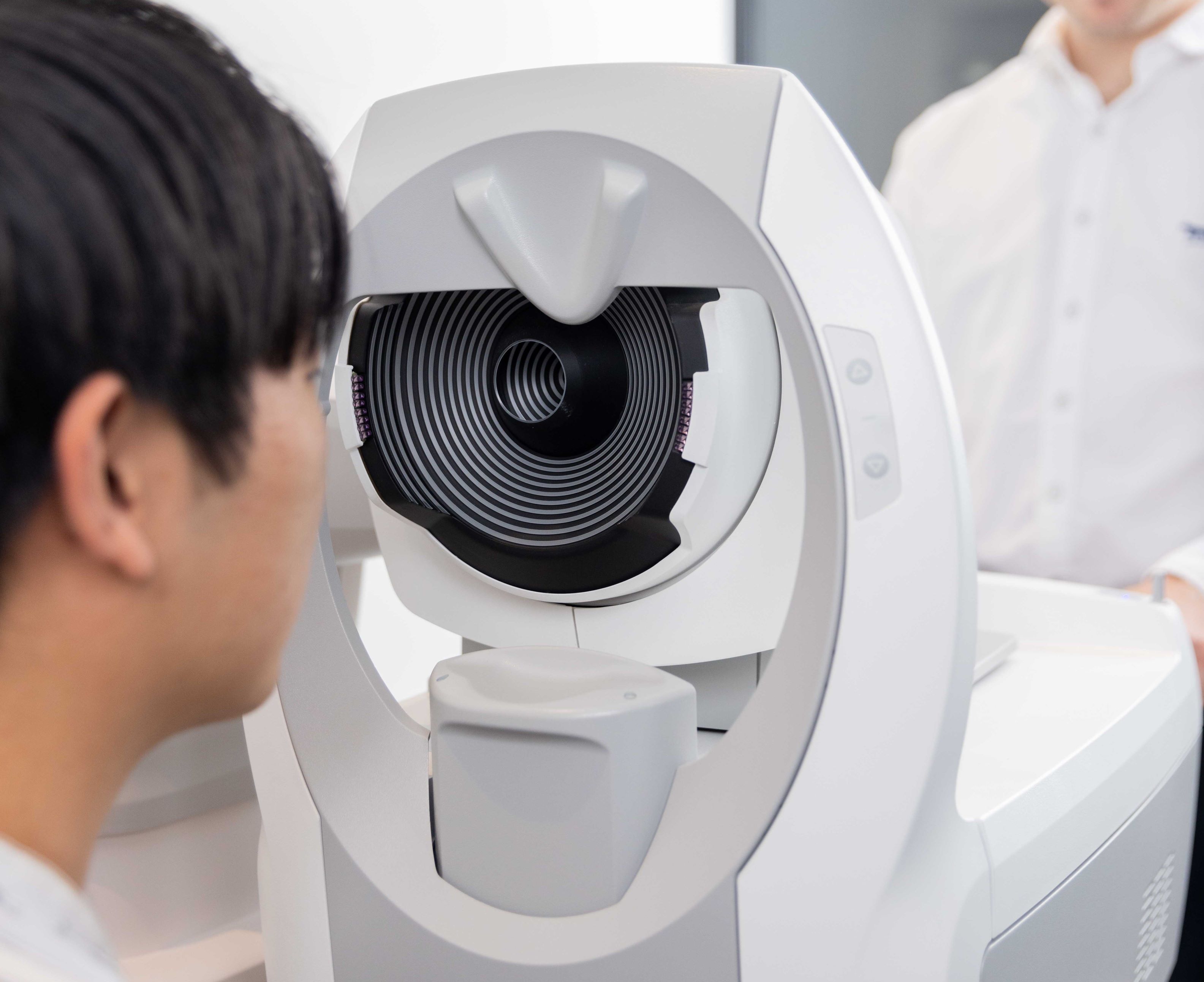
Retinal Imaging
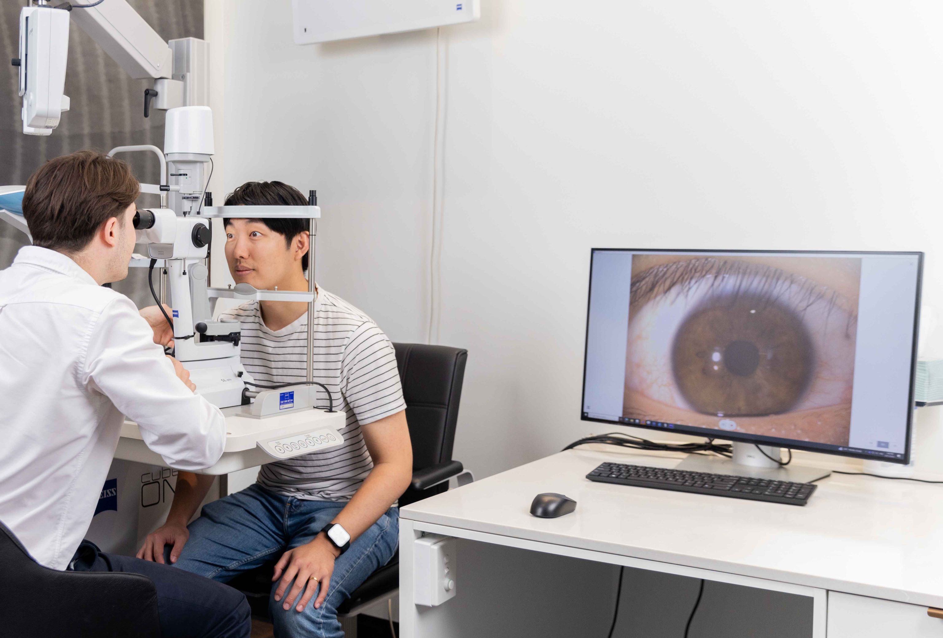
At Innovative Eye Care, your optometrist will take photographs of the back of your eyes using a retinal camera. This process is an important part of your comprehensive eye examination and is used to identify disease and monitor changes to your eye health in the future.
The real value of retinal photography is that it provides a baseline for the condition of your eye, making future changes much easier to monitor. This technology is especially useful in slow-moving pathological processes such as in glaucoma, macular degeneration and diabetic retinopathy. We believe it’s important to have a retinal photograph taken at your initial consultation and then regularly in the future, even if your eyes look normal like the image below. Please be aware that retinal photography does not attract a Medicare rebate so a small extra charge for this service will apply (we are happy to offer 50% off this price for concession cardholders and children).
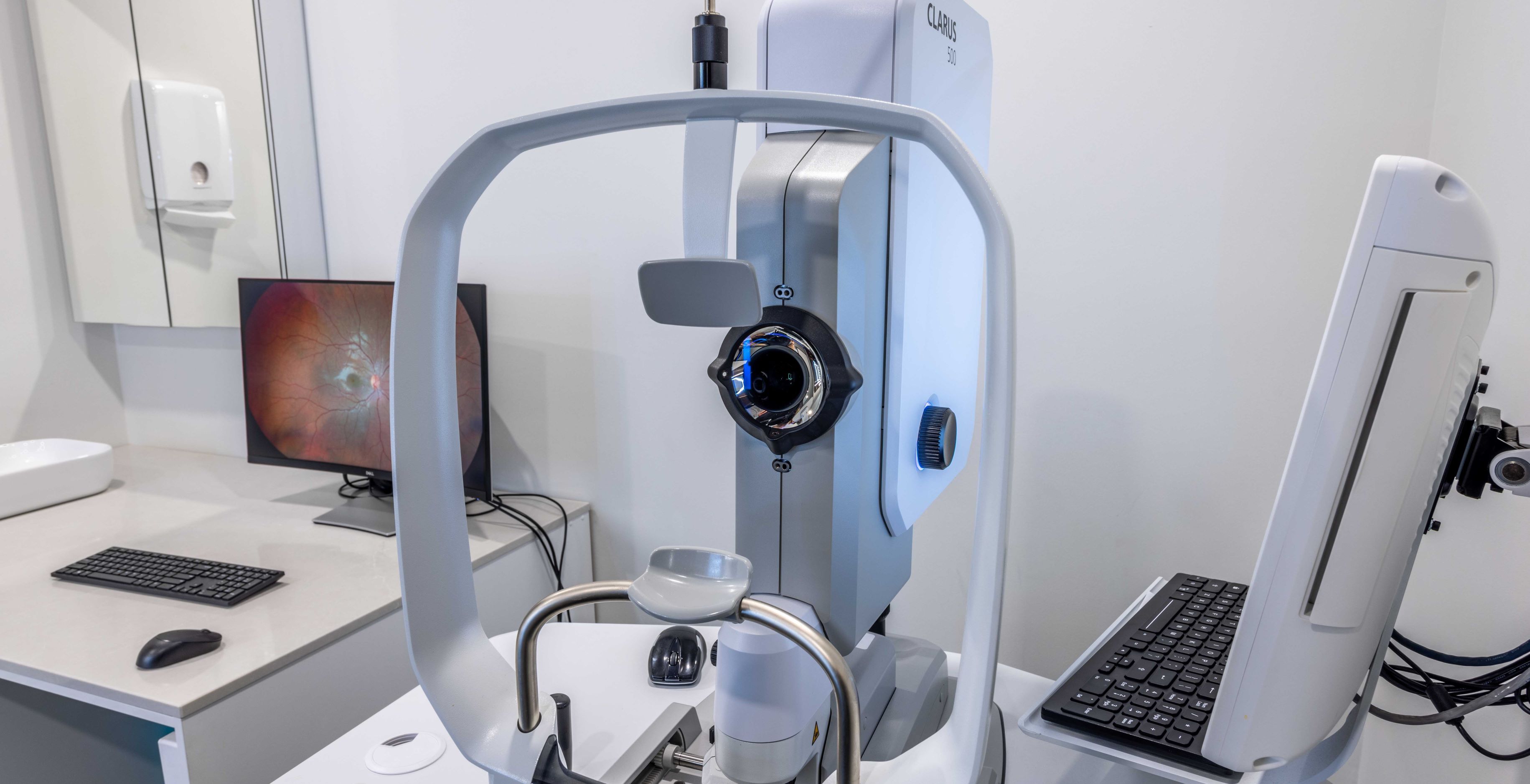
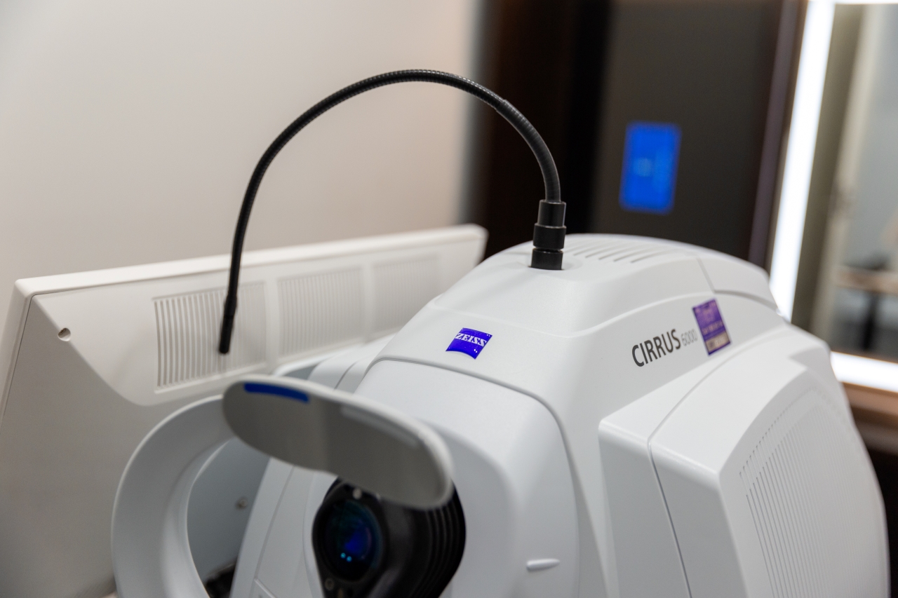
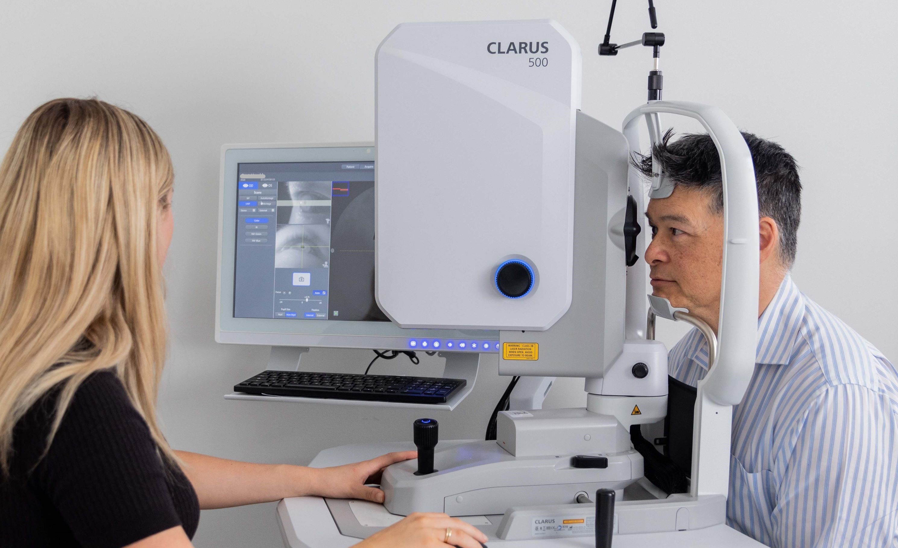
Zeiss Clarus
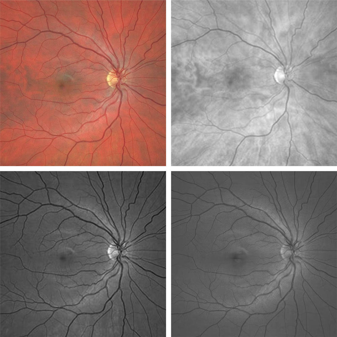
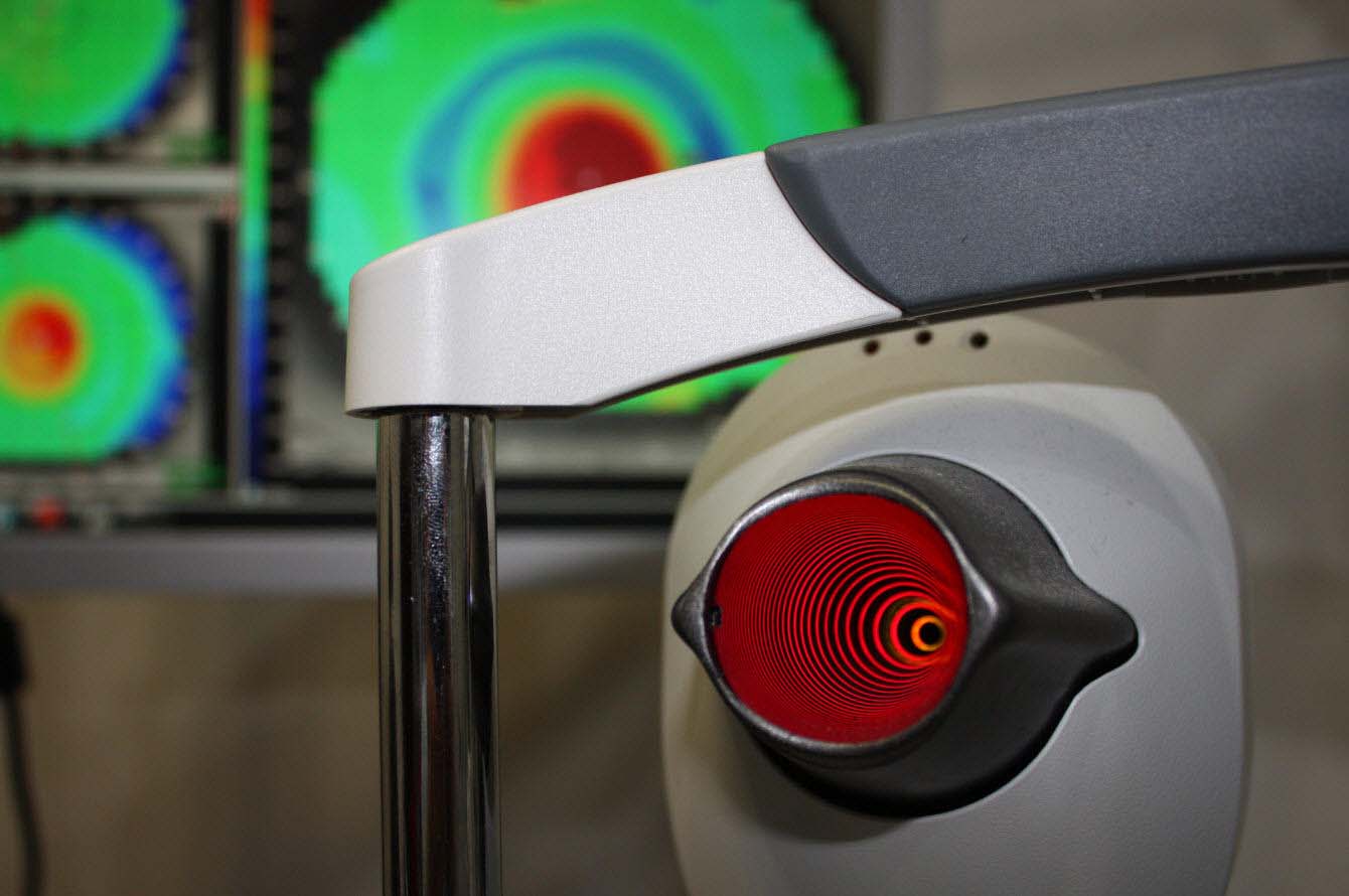
Other imaging modalities often use lasers of only a few specific colours, which can alter the appearance of the retina and make it more difficult to detect any issues. Ultra-widefield photos of the retina are captured with the Zeiss Clarus in true colour to best mimic the actual appearance of the retina. These can be further separated into red, green and blue lasers to show different layers of the retina and any potential diseases located at each; infrared imaging highlights the outermost layer, green imaging highlights blood vessels and blue imaging highlights the nerve pathway.
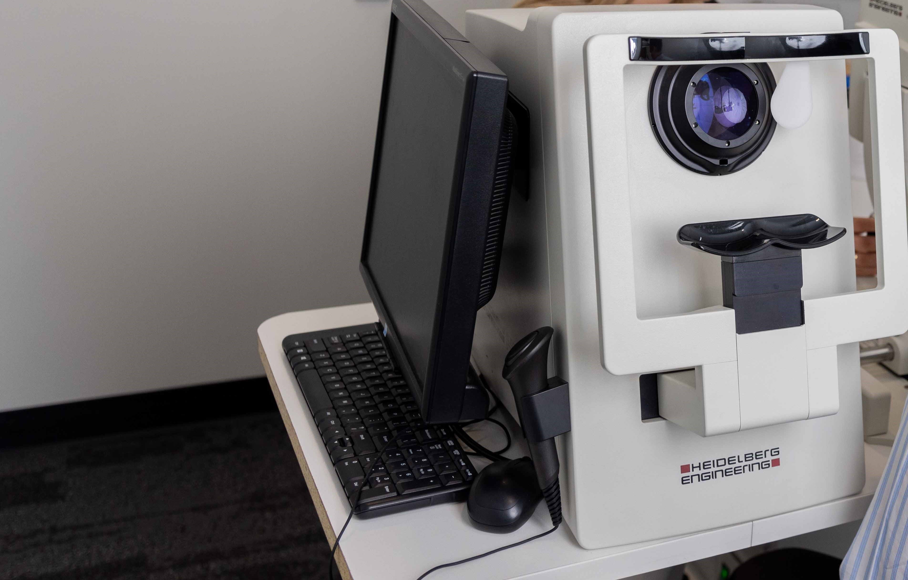
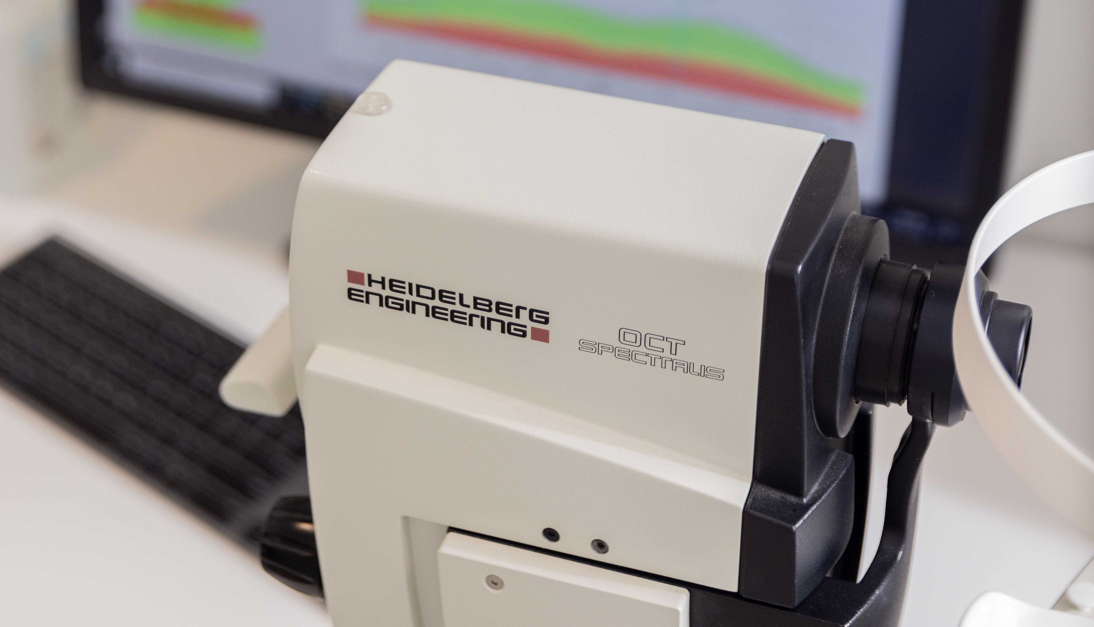
Heidelberg Spectralis
The Heidelberg Spectralis has the ability to take crystal clear photos of the back of the eye by compiling multiple images taken during a 10-20 second scan, capturing an image of the optic nerve, the macula, and the areas beyond. MultiColor photos are also available, using laser imaging on three wavelengths to reveal different layers of the retina.
Both of these types of scans reveal structures and abnormalities of the eye that allow your optometrist to better pinpoint and manage eye health issues, if any, that can be detected using posterior imaging. These methods of diagnostic imaging are offered at our Adelaide practice.

Canon Non-Mydriatic Retinal Camera
Our high-resolution Canon digital retinal camera allows us to take a high-quality photograph of the back of your eye. This allows a more detailed, wider-angled view than would normally be achieved with conventional observation methods, such as a slit lamp, and allows us to store and record visual details of your eye to monitor progression.
FAQs
Please browse through some of our most frequently asked questions on this topic.


Speak to our friendly team today
Book your appointment now for personalised eye care tailored just for you.